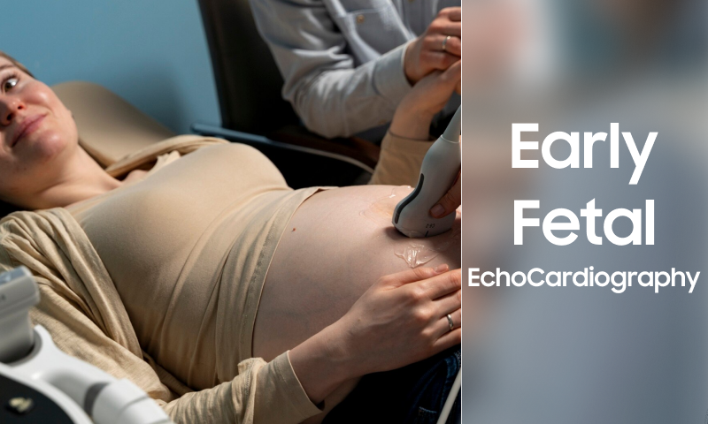
The purpose of early fetal echocardiography, a specialist ultrasound test is to assess the position, size, structure, function, and rhythm of the developing baby’s heart during pregnancy. Also, an obstetrician is simply able to obtain a limited view of the heart of the baby during the regular pregnancy ultrasound. Fetal echocardiography also offers a detailed evaluation of the heart of the baby by experts in fetal echocardiography.
What is the Benefit of early Fetal Echocardiography?
One of the significant benefits of fetal echocardiography is basically the prenatal diagnosis of congenital heart disease, which is also known as CHD. It simply allows the baby to have fast access to medical and surgical intervention after birth. In some of the cases, the prenatal diagnosis is shown to improve the overall outcomes in the babies along with the complex heart disease.
Who Needs a Fetal Echocardiogram?
Some of the women are at a high risk of delivering a baby along with congenital heart diseases. Also, these patients will be considered for the fetal echocardiogram referred.
Indications Include the Following:
- The fetal heart abnormalities are basically suspected from the routine obstetric ultrasound.
- Family history of the CHD.
- Abnormal fetal heart rate or rhythm.
- Abnormality of the other central organ system.
- The twin-to-twin transfusion syndrome.
- Insulin-dependent diabetes mellitus.
- Sjogren’s syndrome or lupus
- Exposure to some medicines in early pregnancy, like some anti-epileptic ones.
- Hydrops.
- Increased nuchal clarity in the first trimester of screening.
- Chromosomal abnormalities are associated with CHD.
Are There Limitations to Early Fetal Echocardiography?
There are some of the abnormalities that a detailed fetal echocardiogram can’t detect prenatally. Also, these will include pulmonary venous anomalies, coarctation of the aorta, small holes, and mild valve abnormalities. Also, some of the cardiac lesions are not evident until the baby is born. Repeat evaluation is required occasionally.

What Information Is Necessary for a Patient to Get Ready for a Fetal Echocardiogram?
Two weeks before the appointment, the patient must receive the appointment confirmation and directions by mail. It is common to request that moms who are less than 24 weeks pregnant attend the doctor with a somewhat full bladder. The fetal echocardiography lasts for one hour as well.
When its Recommended to Get a Fetal Echocardiogram What Takes Place Next?
In terms of scheduling a fetal echocardiogram, a fax referral form needs to be filled and faxed to the heart center at Nationwide Children’s. Then, a representative will simply schedule the urgent fetal test and then return the form to the obstetrician’s office within one working day, along with the appointment date and time.
Generally, the test will be scheduled between 20-24 weeks gestation. if the pregnancy is longer than 24 weeks then the test will be arranged.
When Will I Receive Results?
When the pediatric cardiac sonographer performs the test then, a pediatric cardiologist will simply meet up with the family on the same day to review the results. Also, if the baby doesn’t have any congenital heart disease, then the doctor will discuss the treatment options and prognosis. A plan will also be established for every patient, and follow-up testing will be scheduled when required. The referring physician will also get faxed an overall report on the same day as the fetal echocardiogram.
What If The Baby Requires Special Care?
Suppose it is indicated on the fetal echocardiogram that the baby needs services at Nationwide Children’s Hospital after the birth. In that case, the cardiologist will simply contact the program coordinator of the fetal diagnostics program. Also, the nurse coordinator will simply work closely with the families during diagnosis by helping prepare and educate them about the birth and care of their child. The coordinator also helps to facilitate communication between the health care team and the pediatric specialist as well. The aim is mainly for the families to experience the seamless transition from pregnancy to newborn care simply.
Also, Read – 5 weeks pregnant ultrasound
Final Verdict
So, these are all the details about early fetal echocardiography. We hope that this article helps you to know all the information about it, and if yes, then share this article with others so that they can also understand it. If you need any assistance, you can connect with us by simply dropping a comment in the comment section below.
What is Fetal Echo in Early Pregnancy?
Using sound waves, a fetal echocardiogram can simply make images of the unborn child’s heart. It is a painless ultrasound test that simply shows the structure of the heart and also shows you how well it is working.
How Early Can Fetal Echocardiogram Be Done?
The traditional fetal echo is mainly performed in pregnancies around 20 weeks when the fetal heart is about the size of a quarter. A first-trimester fetal echo is also performed earlier, mainly in pregnancies between 11 and 14 weeks when the fetal heart is about the size of the pea.
Who Needs a Fetal Echocardiogram?
Suppose the month is already having a child born with a heart issue if this is the family history of the genetic heart issue. Suppose the mother has a health condition like diabetes, autoimmune disorders, genetic conditions, or also exposure to some medications.
Does Every Bay Get a Fetal Echo?
Most of the unborn babies do’ n’t need any further testing. The situations when a fetal echocardiogram is required include the following: there is a sibling who was born with a congenital heart defect.
What are the fetal echocardiogram’s limitations?
Certain abnormalities are too minute to get picked up on during pregnancy, even with detailed fetal echocardiography. These comprise mild valve defects, coarctation of the aorta, tiny holes, and anomalies of the pulmonary veins. Furthermore, some heart abnormalities do not become visible until after delivery has happened.
What time is best for doing fetal echocardiography?
Because of the fetus’s small size, imaging the heart becomes increasingly challenging before 18 weeks of pregnancy. So fetal echoes should ideally be done between 18 and 24 weeks of gestation, or roughly in the middle of the pregnancy. But after this particular period, they can be carried out at any time.
